Mitosis Cell Prophase Metaphase Anaphase Telophase Biology Diagrams The following points highlight the six main stages of mitosis cell division. The stages are: 1. The Interphase Stage 2. The Early Prophase Stage 3. The Late Prophase Stage 4. The Metaphase Stage 5. The Anaphase Stage 6. The Telophase Stage. 1. The Interphase Stage: The slide shows Interphase stage which is characterized by the following

As living organisms grow, their cells must replicate and divide. Most animal cells, except for sex cells, undergo the process of mitosis to create new cells. Through mitosis, one cell creates two genetically identical daughter cells. Mitosis is a complex process consisting of multiple phases; anaphase, interphase, metaphase and prophase. Each phase has its own steps and is integral to the

Edexcel The stages of mitosis in detail Biology Diagrams
Interphase The cell spends most of its life in this phase. The DNA in chromosomes copies itself ready for mitosis. Prophase The DNA in chromosomes and their copies condenses to become more visible

Learn the four stages of mitosis, the process of cell division that produces identical daughter cells. Prophase is when chromatin condenses into chromosomes and the spindle apparatus forms.
Stages of Mitosis Cell Division Biology Diagrams
Some cells remain in interphase for days or even years; some cells never leave interphase. At the end of interphase, the cell has duplicated its chromosomes and is ready to move them into separate cells, called daughter cells. This occurs during the four steps of mitosis, called prophase, metaphase, anaphase and telophase.
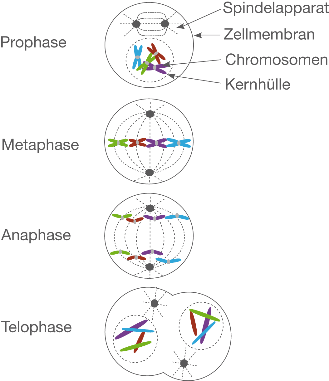
Prophase. Most of the time, DNA is tightly coiled and structured around proteins called histones. This packaged form is known as chromatin. The first stage of mitosis sees this chromatin supercoiling from their operational width of 30nm, to the 500nm thickness associated with chromosomes.
